Freitag, 09. M�rz 2018, 15:30 - 17:00 iCal
Optical nanoscopy: J.G. DANZL (IST Austria)
"Optical nanoscopy – bridging scales from molecules to tissues"
Gastvortrag von Johann G. DANZL (IST Austria) im Seminar für Physikalische Chemie und Materialchemie
Chemische Institute, Hörsaal 4, Halbstock
Währinger Straße 42, 1090 Wien
Seminar, Workshop, Kurs
Far-field (lens-based) light microscopy is a central tool in a diverse range of research areas. In the life sciences, it con-veniently allows the observation of specific molecular entities and - as fluorescence microscopy - enables high detection efficiency, down to the single molecule level. Most strikingly, it allows decoding the structure and dynamics of living cells and tissues in a minimally invasive way.
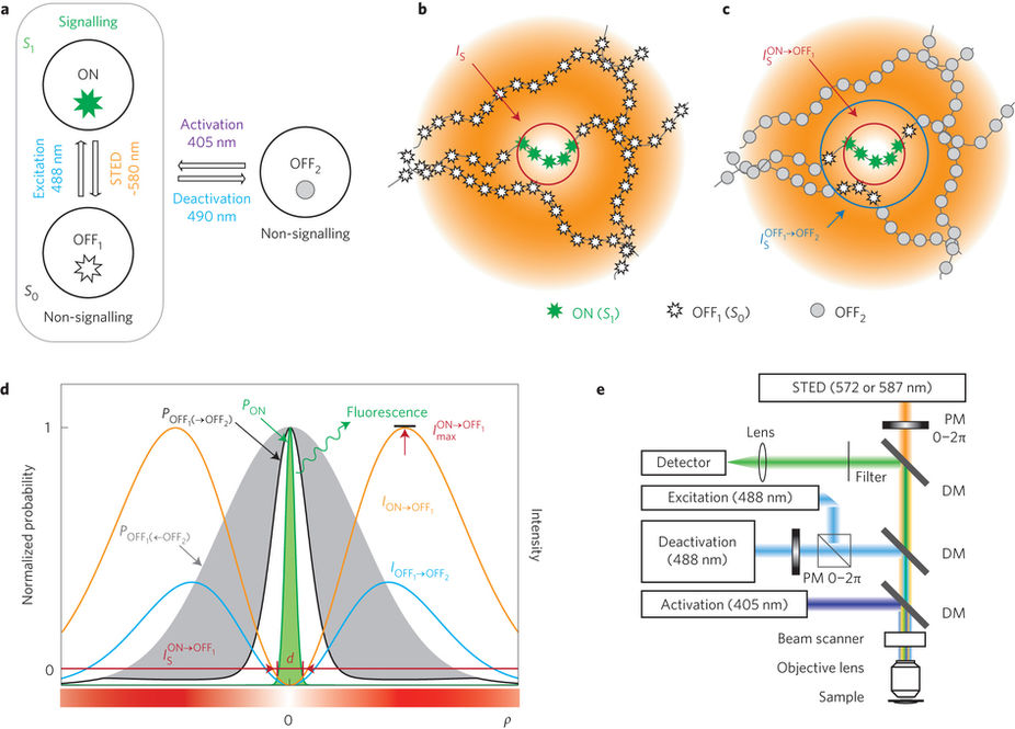
However, diffraction of light waves poses a limit to the attainable spatial reso¬lution on the order of half the wavelength of visible light or 200 nm. Recently, several powerful methods termed “super-resolution” optical imaging or “nanoscopy” have been developed that enable resolution better than the diffraction limit, in the range of tens of nanometers. I will give an introduction to diffraction-unlimited optical imaging with an emphasis on the so-called “coordinate-targeted” nanoscopy methods, where a pattern of light determines the sub-diffraction regions where fluorescence is allowed to originate from, thus improving resolution.
Unraveling details of the biological specimen much finer than the diffraction resolution limit in living cells still poses a con-siderable challenge. This is particularly true when dynamical changes or the three-dimensional (3D) structure of the living system are to be deciphered at high resolution. I will give a specific example of how to improve these coordinatetargeted methods. By using two, rather than a single, molecular off-transition to determine the regions of allowed fluorescence, we achieved a significantly improved repeated imaging capability and improved state contrast and resolution. This enabled e.g. decoding the 3D structure of neurons in living brain tissue, as exemplified in the image to the left. Here, a portion of a nerve cell is shown as a 3D rendering with the actin cytoskeleton labelled. A multitude of so-called dendritic spines emanate from the dendritic shaft (Danzl, Sidenstein et al., 2016, Nature Photonics 10:122). The spines are the location of the synapses, where information is passed from one neuron to the next and that play a central role in the brain’s capability to learn and form memories. Furthermore, I will give an outlook on current research efforts in my laboratory.
ist.ac.at/research/research-groups/danzl-group/
Veranstalter
Institut für Physikalische Chemie, Universität Wien
Kontakt
Univ.-Prof. Dr. Wolfgang Kautek
Universität Wien
Institut für Physikalische Chemie
0043 664 60277 52470
wolfgang.kautek@univie.ac.at
Erstellt am Dienstag, 06. Februar 2018, 11:59
Letzte Änderung am Mittwoch, 07. Februar 2018, 08:43